The abdominal wall forms the anterior and lateral parts of the abdominal cavity and consists of multiple layers built on top of one another. Understanding the structure of the abdominal wall is crucial for understanding how hernias develop and how they are treated.
Layers of the abdominal wall
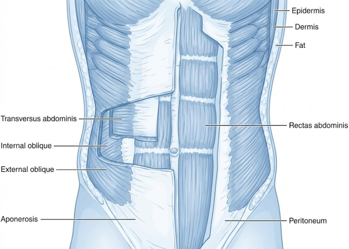
Proceeding from outside to inside, the outermost layer is the skin, beneath which lies a subcutaneous connective tissue layer that also contains fatty tissue. Beneath this are the abdominal muscles, which consist of strong, flat muscle sheets.
At the front, the best-known muscle is the rectus abdominis, which runs vertically from the chest to the pubic bone and is divided in the midline by a tendinous band called the linea alba (white line). On the sides, three muscle layers are arranged one above the other: the external oblique, the internal oblique, and the transverse abdominis. These run in different directions, providing a high degree of stability and freedom of movement.
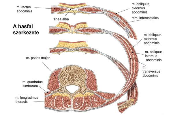
The abdominal muscles are covered by connective tissue sheets (fasciae), which assist in force transmission and hold the muscles together. The innermost layer is the parietal peritoneum, a thin membrane that separates the abdominal wall from the internal organs.
The function of the abdominal wall
The abdominal wall serves to protect the organs of the abdominal cavity, maintain intra-abdominal pressure, and participate in trunk movement, posture, and everyday processes such as coughing, laughing, and defecation.